
Results#
The continuous evolution campaign has not been run. This page describes the designed shell and circuit, the completed assays and the outlook.
| Subsystem | State |
|---|---|
| Shell variants (inside engineering) | Cloned and sequence-verified (p_f008–p_f011) |
| Shell expression in E. coli BL21 (DE3) | Confirmed by SDS-PAGE and anti-His immunoblot |
| MutaT7 mutagenesis | Functional (stop-codon reversion assay) |
Mutation plasmids (p_m005–p_m007) |
Cloned and sequence-verified |
| Guide truncation series | Designed: non-targeting, 10, 11, 14, 17, 20 nt |
Selection plasmids (p_s002 series) |
Assembly in progress, see 3.6 |
| Cage assembly and co-encapsulation | Not yet demonstrated, see 3.4 |
| Continuous evolution | Planned |
3.1 Engineering QtEncapsulin for protein–RNA co-encapsulation#
The cargo pair is the ribonucleoprotein of catalytically dead Cas9 (dCas9) and its sgRNA. The two halves are chemically distinct and each gets its own handle: dCas9 through a fused cargo-loading peptide (CLP), the sgRNA through a boxB hairpin.
Protein handle. The CLP core motif is five residues and docks to a site on the lumenal face of the shell, so fusing it is sufficient to direct a protein inside.1 The IMEF targeting peptide is attached to dCas9 at the established C-terminal fusion site.
RNA handle. QtEnc has no native nucleic-acid affinity. The arginine-rich λN⁺ peptide binds the boxB stem-loop with high affinity and is small enough to graft.2 Terasaka et al. used cationic peptides including λN⁺ to give a non-viral cage mRNA recognition, with the peptides lining the lumenal edge of the shell.3 λN⁺ is therefore displayed on the lumenal face of QtEnc. In the sgRNA, boxB motifs replace the validated MS2 stem-loop insertion sites in the scaffold stem-loop and tetraloop (Fig S2).
External modifications. The outer shell was first given a His-tag, for purification and immunoblotting, and a targeting peptide (TP), for delivery. That combination produced insoluble material (see 3.4). Which of the two is responsible was not resolved, so the designs used from there on carry no external modification. Parallel designs retaining the TP without the His-tag were also built, to test the TP's effect on its own.
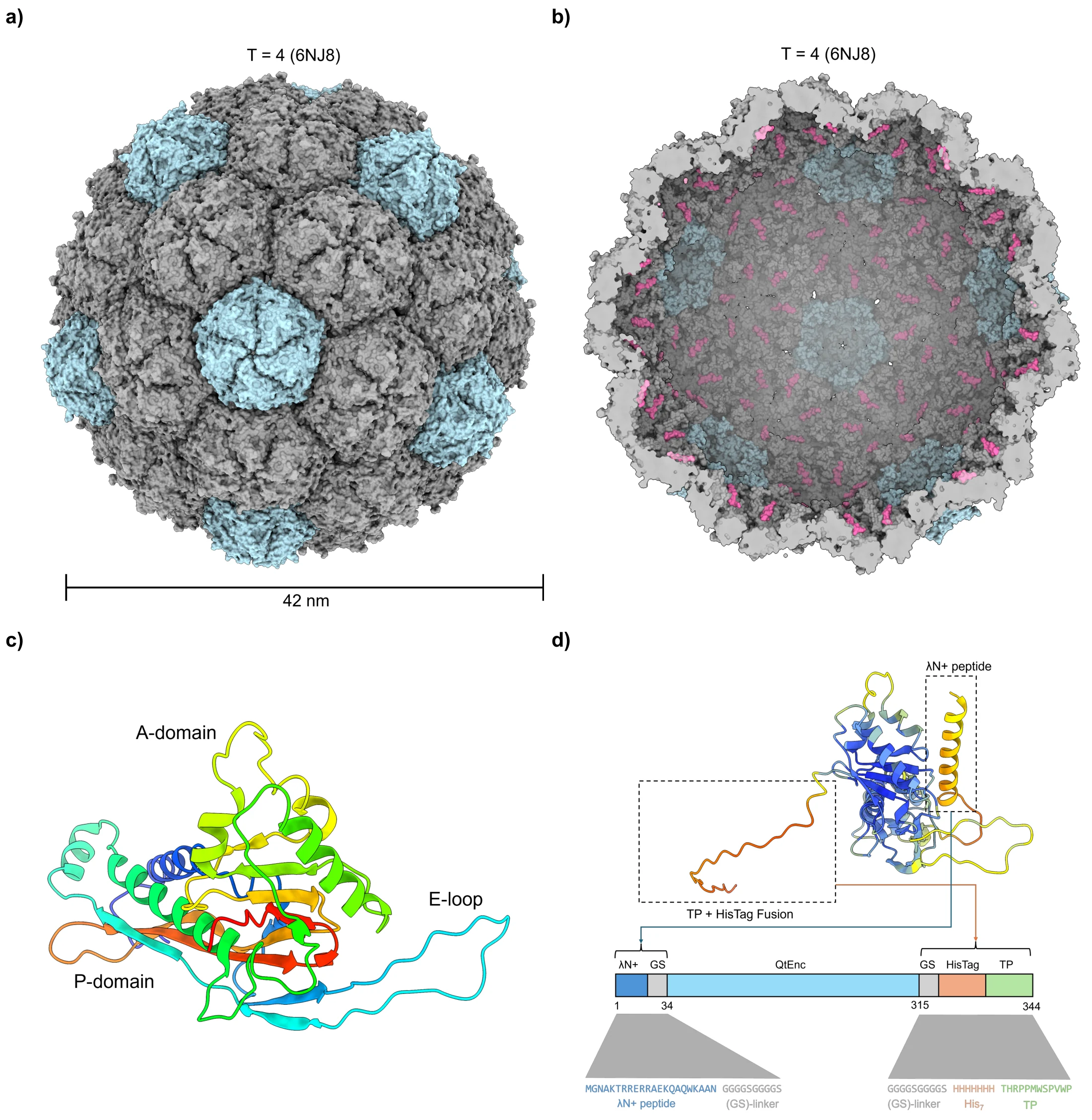
Fig 1. Structural overview of T = 4 QtEnc (6NJ8). (a) Outer shell viewed down the 5-fold axis. (b) Cross-section with the IMEF targeting peptides (TP) in pink. (c) A single subunit in rainbow colouring, with the A-domain, P-domain and E-loop marked. (d) AlphaFold 3 model of QtEnc-HisTag-TP-λN⁺ coloured by pLDDT; the HisTag and TP sit outside and λN⁺ inside, each joined to QtEnc by a GS linker, with the sequence map below.
3.2 An encapsulation-coupled selection#
Engineering inside and outside the shell introduces several properties that can be improved by evolution: assembly (efficiency, monodispersity, T-number), stability, and function (protein and RNA loading, tolerance of surface display). The selection rewards the last two, through the first.
A dCas9–sgRNA complex represses a kanamycin-resistance gene (kanR) on the selection plasmid. The proposed circuit targets the resistance ORF to produce graded repression. Capturing dCas9 or its sgRNA inside QtEnc is intended to relieve repression and restore growth (Fig 2). Vigouroux et al. showed that the level of complementarity between guide and target sets repression in defined steps, with less variation than titrating dCas9 itself.4 The guide truncation series (10, 11, 14, 17 and 20 nt, plus a non-targeting control) turns that into a tunable selection pressure (Fig 3). The mechanism page gives the kinetics.
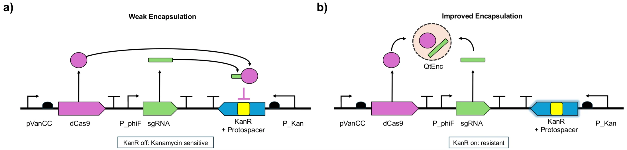
Fig 2. KanR-dependent positive selection. (a) Weak or absent encapsulation: free dCas9 and sgRNA form an active complex that binds the protospacer in the resistance-gene body, reduces transcription and leaves KanR off in this schematic: the cell is kanamycin-sensitive. (b) Improved encapsulation: evolved QtEnc variants capture dCas9 or its sgRNA, reducing the free repressor complex. Repression is relieved, KanR is on, and the cell has a selective advantage. Growth rescue alone does not demonstrate capture of both components.
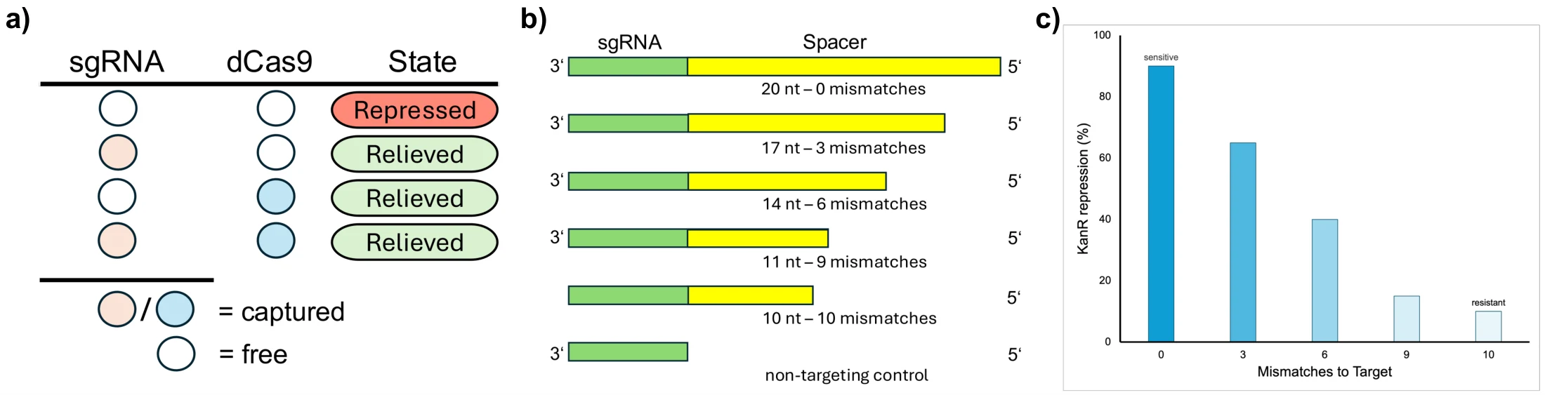
Fig 3. Principle, design and tuning of the selection. (a) When both components are free in the cytoplasm they repress KanR (Repressed); if at least one is enclosed in the capsid (Captured), repression is relieved (Relieved). (b) sgRNA variants for tuning binding affinity: the spacer is progressively truncated while the scaffold, including the PAM-proximal region, stays intact, alongside a non-targeting control. (c) Repression of KanR as a function of spacer complementarity. More truncation means more mismatches, weaker binding and less repression, so stringency can be set between sensitive and resistant.
Regulation and an alternative readout#
dCas9 and the sgRNA are expressed under the VanRAM and PhlFAM regulators, which Meyer et al. found to have the largest dynamic range of a dozen tested while staying orthogonal to each other.5 Guide complementarity and inducer concentration are then two independent axes for tuning the selection, and the pressure can be changed over time.
A second readout based on the CRISPR-regulated toxin–antitoxin system creTA was explored as an alternative to kanR. In archaea, the creA RNA represses creT, a small RNA toxin that sequesters rare codons.6 Chen et al. built a sensitive positive selection on creT for evolving Cas12a.7 Placing the sgRNA's protospacer in the 5′ UTR of the repressor that controls creT gives the same logic: encapsulating dCas9 or the sgRNA is rewarded with growth (Fig S1).
3.3 Mutation and selection plasmids#
The circuit is split across two compatible plasmids so that only the shell gene is diversified. The mutation plasmid carries the MutaT7 fusion and the QtEnc open reading frame between a T7 promoter and terminator; that cassette is the only hypermutated locus. The selection plasmid carries dCas9, the sgRNA cassette and kanR (Fig S3).
The split is a containment measure. A mutation that lowers dCas9 or sgRNA expression restores resistance without improving encapsulation and would be enriched as strongly as a genuine improvement, so both sit outside the T7 transcription unit. That protects them from hypermutation, not from host polymerase error, so the selection plasmid is re-sequenced periodically during passaging.
3.4 Purification and validation of QtEncapsulin cages#
His-tagged QtEnc (QtEnc-His) was expressed in E. coli BL21 (DE3). Monomer expression was confirmed by SDS-PAGE across the purification steps (Fig S11), and the monomer elutes at 16–17 mL on Superose 6 (Fig S7, S12).
The His-tag moves QtEnc into the insoluble fraction. After Ni-NTA purification of the soluble fraction, analytical SEC shows no void-volume peak (Fig S8). Fractionating the lysate shows that most QtEnc-His is in the pellet (Fig 4e). Continuing from the insoluble fraction by heat precipitation, without Ni-NTA, gives a void-volume peak in SEC (Fig 4a) and a high-molecular-weight species on Blue Native PAGE that reacts with anti-His (Fig 4d, Fig S10). Of the four workflows compared, only the pellet-derived, heat-precipitated sample shows this peak (Fig S4).
Size by DLS. Fractions across the QtEnc-His void-volume peak read 209–340 nm (Fig 4c), against 42 nm expected for a T = 4 cage, so the species is larger than a single cage. Void-volume elution and native-gel migration cannot separate large aggregates from assembled cages; negative-stain TEM or cryo-EM is the orthogonal test (see outlook). Removing the His-tag recovers QtEnc in the soluble fraction, with a void-volume peak at 7.6 mL (Fig 4b). DLS of wild-type QtEnc and of QtEnc-TP-mScarlet from the crude Sephacryl S-500 step gave no defined peak near 42 nm, and the intensity and volume distributions differ in a way that indicates aggregation (Fig S5, S6).
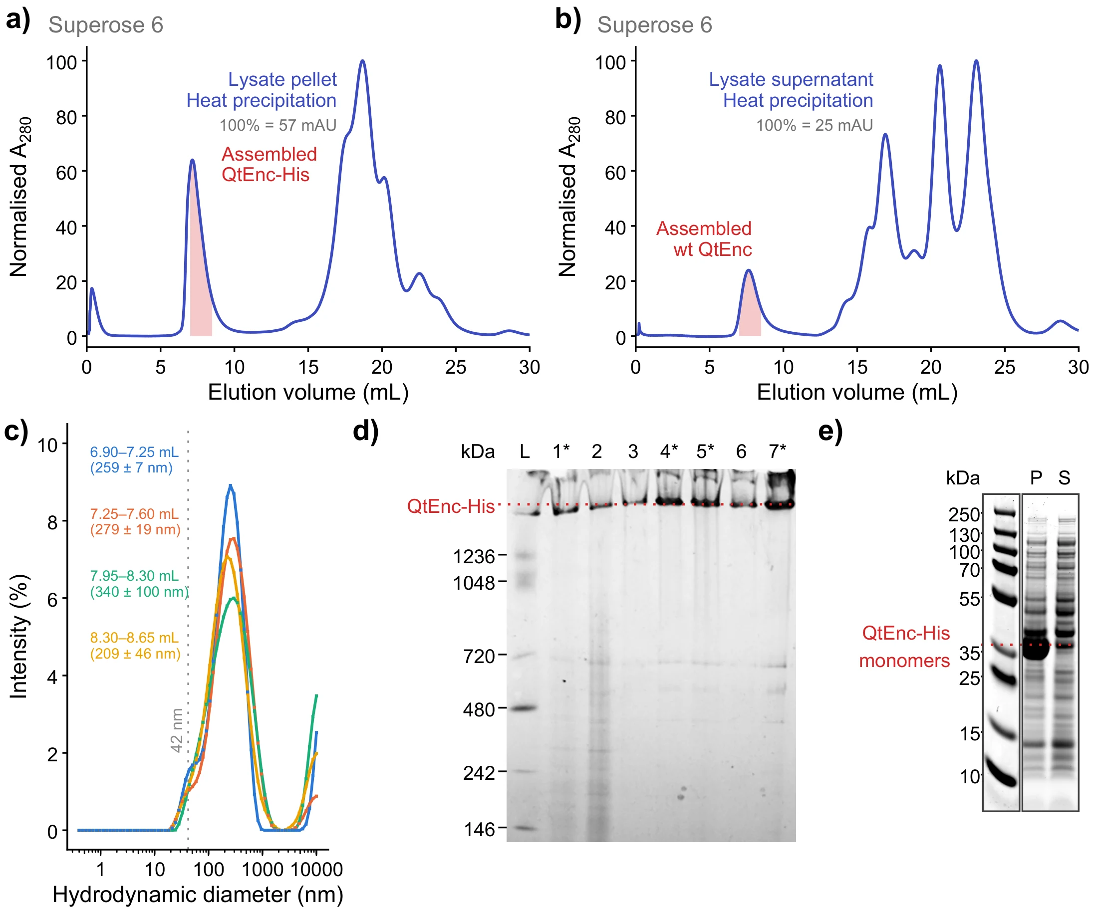
Fig 4. Analytical SEC after heat precipitation on a Superose 6 10/300 GL column. (a) QtEnc-His from the insoluble lysate fraction (resuspended pellet), purified without Ni-NTA, and (b) wt QtEnc from the clarified lysate supernatant. Both show a peak near the void volume, at 7.2 mL in a and 7.6 mL in b (shaded, 7–8.5 mL). A280 is normalised to the maximum of each trace after 1 mL (100 % = 57 mAU in a, 25 mAU in b). (c) Intensity-weighted DLS of four consecutive 0.35 mL fractions across the void-volume peak in a, each the mean of 2–3 runs, labelled by elution volume; brackets give the peak diameter as mean ± SD across runs. The dotted line marks the 42 nm expected for the T = 4 cage; every fraction peaks between 209 and 340 nm. (d) Blue Native PAGE across the QtEnc-His purification by heat precipitation, Coomassie-stained. L, NativeMark standard; 1, pellet after lysis; 2, supernatant after lysis; 3, pellet after heat precipitation; 4, supernatant after heat precipitation; 5, supernatant after filtration; 6, Amicon flow-through; 7, concentrated sample. Asterisks mark the fraction carried forward at each step (lanes 1, 4, 5, 7). QtEnc-His migrates above the 1236 kDa standard. (e) SDS-PAGE of the pellet (P) and supernatant (S) after lysis: most QtEnc-His is in the pellet.
3.5 Stop-codon reversion assay#
Before any evolution campaign, MutaT7 mutagenesis was tested in a simple reporter. An early stop codon was placed in a chloramphenicol-resistance gene (CmR) by changing a Trp codon to a stop (TGG → TAG), so restoring resistance needs the A→G deamination activity of MutaT7. Colonies grew on 25 µg/mL chloramphenicol (Fig S13), so the system is operable.
The stop codon reverted under all three conditions (+IPTG, +glucose and basal) with similar colony counts for the undiluted cultures. That points to leaky MutaT7 expression, which the outlook addresses.
3.6 Cloning of the mutation and selection plasmids#
Constructs were assembled by Golden Gate with synthetic gene fragments (Twist Bioscience) and PCR products. All mutation plasmids were cloned and sequence-verified. For the selection plasmids, whole-plasmid sequencing of the Golden Gate reactions shows that four of the five BsaI junctions form and that all five parts are covered without gaps (Table S2). No read spans all junctions at once, so assembly of the full plasmid is the open step. The plasmid inventory is in Table S1.
Outlook#
The campaign depends on three steps, listed in the order in which they can be tested.
1. Cage assembly with and without the external tag. The data indicate that the His-tag causes aggregation. The next constructs drop it and keep the targeting peptide, and a DLS baseline on wild-type cages gives the reference for 42 nm. Negative-stain TEM or cryo-EM would confirm assembly independently of elution volume and native-gel migration. Mixing monomers with and without an external moiety in defined ratios, to make mosaic cages, is a further route to tolerating external fusions.8
2. The selection plasmids. The J5 junction (TcR to backbone) is the one not
observed: the backbone ligated to s001 through an overhang pair differing at a
single position (GATA/GAAA), excising the TcR cassette. That explains the
missing cassette but not the lack of colonies, since the product still carries
the kanamycin marker. Leaky expression of dCas9 and the guide, unrepressed
without VanRAM and PhlFAM, is the more likely burden. The
options are chemical promoter repression, a non-targeting guide as a proof of
concept, and a tri-plasmid design that introduces the regulators first, from a
separate plasmid.
3. Mutagenesis control. An inducible sgRNA directed at the T7 promoter region would suppress the leaky MutaT7 activity seen in 3.5.
With these in place the planned campaign can start, and enriched shell alleles can be re-cloned and tested by the co-encapsulation assays. The longer list is on the future work page.
-
Cassidy-Amstutz et al., Biochemistry 55, 3461 (2016); Giessen et al., eLife 8, e46070 (2019). ↩
-
Lazinski, Grzadzielska & Das, Cell 59, 207 (1989); Baron-Benhamou et al., Methods Mol. Biol. 257, 135 (2004). ↩
-
Terasaka et al., Proc. Natl. Acad. Sci. USA 115, 5432 (2018). ↩
-
Vigouroux et al., Mol. Syst. Biol. 14, e7899 (2018), doi:10.15252/msb.20177899. ↩
-
Meyer et al., Nat. Chem. Biol. 15, 196 (2019). ↩
-
Li et al., Science 372 (2021), "Toxin–antitoxin RNA pairs safeguard CRISPR-Cas systems". ↩
-
Chen et al., Adv. Sci. 12, e17105 (2025). ↩
-
Boyton et al., ACS Omega 7, 823 (2022); Charron et al., FEBS J. 293, 1908 (2026). Full list on the references page. ↩